We are sort of eye nerds around here…
Optometry has moved forward in leaps and bounds over the 40+ years we’ve been here and we are constantly adding the latest in technological advancements to our toolkit to help us diagnose and improve your most important sense.


Phoropter
The Phoropter is a very precise Ophthalmic Testing piece of equipment that lets us test over 13,000,000,000,000,000 (quadrillion) combinations of lenses in front of your very eyes.
We’ll try out various combinations of lens attributes while you look through the instrument at an eye chart across the room. This lets us find the best possible correction for your eyes by effectively letting you try on all the options we think will work best.

Ultra Wide Field Internal Imaging Camera
The Ultra Wide Field Internal Imaging camera takes pictures of the retina (the inside of your eye). This advanced instrument allows us to take an incredible 200 degree wide picture which lets us see and detect more problems.
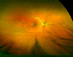 Many eye problems as well as health problems can go undetected for years. Whether these diseases are macular degeneration, retinal detachments, retinal tears, glaucoma, or health issues like high blood pressure or diabetes, these can be determined through the ultra wide field internal imaging.

Automated Refractor
The automated refractor lets us objectively measure the ability of your eyes to focus light correctly on your retinas.
 While you watch a picture move in and out of focus, a computer records the way the image appears on your retinas. By evaluating this image, we obtain a map of the curves of your cornea and use this information to determine a starting point for the prescription of your glasses or contact lenses.
The refractor helps us determine if your eye lids are impacting your sight and precisely measures the distance between your eyes which is paramount in designing your glasses.
By recording and comparing these images over time, we can monitor corneal changes and the progression of various optical diseases.
new-tonometer")
Tonometer
A tonometer measures the pressure inside of your eye with a puff of air.
Our very special advanced tonometer not only measures this with a softer puff of air, but it also automatically corrects the pressure readings by taking into account the corneal thickness and strength. This gives us a more accurate measurement and helps the Doctor understand the health of your eyes.
Knowing the pressure inside your eye, and how it changes over time, helps us monitor your eye health in special conditions like glaucoma, Fuchs’ dystrophy, post-LASIK, corneal edema, refractive surgery, keratoconus, and irregular or thin corneas.

Slit Camera
The slit lamp gives us a magnified, three-dimensional view of your eye. We can use the slit lamp to look at the front parts of the eye (cornea and sclera), the lens, the colored part (iris), and the front section of the gel-like fluid that fills the large space in the middle of the eye.
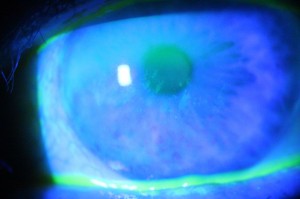 Special lenses can be placed between the slit lamp and the cornea to view the deeper structures of your eyes, such as the optic nerve, retina, and the area where fluid drains out of the eye.
The slit lamp helps us detect many disorders throughout the eye, identify foreign objects in the eye and detect eye problems that may be caused by other diseases or contact lenses.

Eyescan
Our eyescan uses light which reflects off the tissues of the eye to provide a three-dimensional (3D) cross-section of the inside in microscopic resolution. Just like an ultrasound but with light!
This huge leap in technology allows us to better see and diagnose changes in your eyes. The key to preserving your sight is through early detection, so this technology is giving you the best chance to maintain healthy eyes. The best part is the EyeScan is painless, non-invasive, non-contact and only uses light!
This is especially important for diagnosis of macular degeneration, monitoring glaucoma, diabetic eye diseases, other blood-related diseases and detecting neurological diseases.

Healthy Retina

Macular Degeneration

Macular Pigment Optical Density Measurer
A unique instrument that measures your Macular Pigment. Macular pigment measurement allows us to asses a key risk factor in age-related macular degeneration.
This is an important biomarker that relates to your ability to maintain the health of the tissue by minimizing the cumulative damage from ultraviolet radiation, high-energy visible light, and natural cellular degeneration. It also maintains visual performance in settings for glare, bright light, and night driving.
We believe that having the right tool for the job is important. This is an important way to quantify a modifiable factor for you. Like blood pressure or eye pressure.
This instrument is the only technology scientifically proven to measure this modifiable factor which can be treated with vitamin therapy.

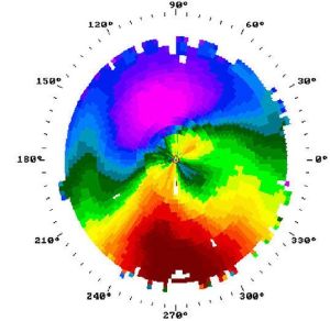
Visual Field Tester
A visual field test checks the extent and intensity of what you are able to see including your entire peripheral (side) vision. By measuring the extent and the intensity of one’s visual field, many visual and general health conditions can be detected.
During the test, you will watch a red light. The computer will randomly flash small lights in your side vision. When this occurs, you push a button to tell the instrument that you were able to see the light. It can tell when you are not looking straight ahead and will retest all the points that you missed.
Some of the things we look for are glaucoma, retinal detachments, retinal degeneration, optic neuritis, brain tumors, toxic effects of ingested items, the cause of visual flashes and floaters, and the visual field effects of traumatic and vascular cerebral and ocular accidents.

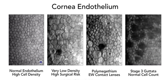
Specular Microscope
We have a very unique and special Specular Microscope that evaluates the health of the cells lining the inner most layer of your cornea. Being the only cells in the body that do not regenerate, depending on the corneal health, these cells change size and shape to fill in the loss of the other cells.
The reason for the loss of cells can be attributed to many different situations. These situations include corneal diseases, contact lens overuse, extended contact lens wear, and intraocular surgeries.
The corneal cell count can allow us to measure and calculate the endothelial cell density of each patient. This can help us determine the correct course of action needed to protect the sight and health of your eyes.

Our infant vision tester makes it possible for us to test and evaluate the sight of very young children. We use the principle of “preferential looking,” or “silent speech,” for testing your baby. The test reveals what an infant can or cannot see.
It is advisable to have all infants checked at 6 months of age by an eye care provider, we specialize in that service.
Scoring is based on observation of the child’s visual response to stimulus targets; and visual acuity or “sharpness” is expressed in standard Snellen measurement as used with older children and adults. i.e. 20/20, 20/40, etc.
We are able to evaluate all the functions that we can with an adult. We can determine eye development and eye health (check for glaucoma, cataracts, congenital or acquired diseases); make sure the muscles and nerves are all present and functioning; that the eyes are working together as a team, that there is no amblyopia (lazy eye) or strabismus (eye turning). We can check for farsightedness, nearsightedness, or astigmatism; and we can evaluate and follow the development of the eyes and their acuity. We do all of these tests by objective procedures, which do not require patient resources.